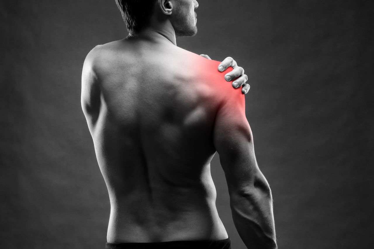
Before we dig into the operating muscles of the shoulder and begin to “dissect” it, it is crucial to understand that the shoulder is built out of 2 joints that form the shoulder as we know it.
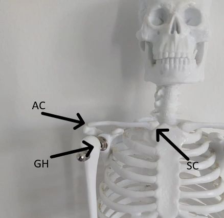
The first joint is the Glenohumeral (GH) Joint. This joint is a ball and socket type that connects our arm (Humerus Bone) to the torso through the Glenoid Cavity of the Scapula.
The second joint is the Acromioclavicular (AC) Joint. This joint is the connection point of our Clavicle and the Scapula through the Acromion process. An extension of this joint is the is the Sternoclavicular (SC) joint, while it is not part of the shoulder joints when looking on the anatomical build of the joint, it is inseparable from the shoulder, as any dysfunction in it would immediately affect the AC joint due to the shared bone of the clavicle.
Now, after we’ve cleared that the shoulder is actually a complex joints structure, we can understand that pain can be caused due to dysfunction on either the joints, as it would affect the rest of them as well.
The shoulder can perform movement in all the movements planes. The movements include flexion and extension of the shoulder on the Sagittal plane, abduction and adduction on both the horizontal and the frontal planes, internal rotation and external rotation along with elevation and depression of the shoulder. These are the movements we are going to discuss and check what could cause pain in any of them.
Each motion can be done through several muscles, but it is also important to note that each muscle might be able to perform several motions. We will examine the optional muscles that could be causing the pain along with the movement they create.
This article contains muscular related pain. A proper examination may rule out muscle related pain, and you will be referred to get a scan to check the structures of the shoulder. This article was created to give some idea of what is going on in the joint when you feel pain, but should not be a replacement for an appointment with a certified therapist or your GP.
If the following information leaves you with an unanswered question, that is completely understandable, feel free to send us a message describing your situation so we could discuss it specifically.
Shoulder Flexion
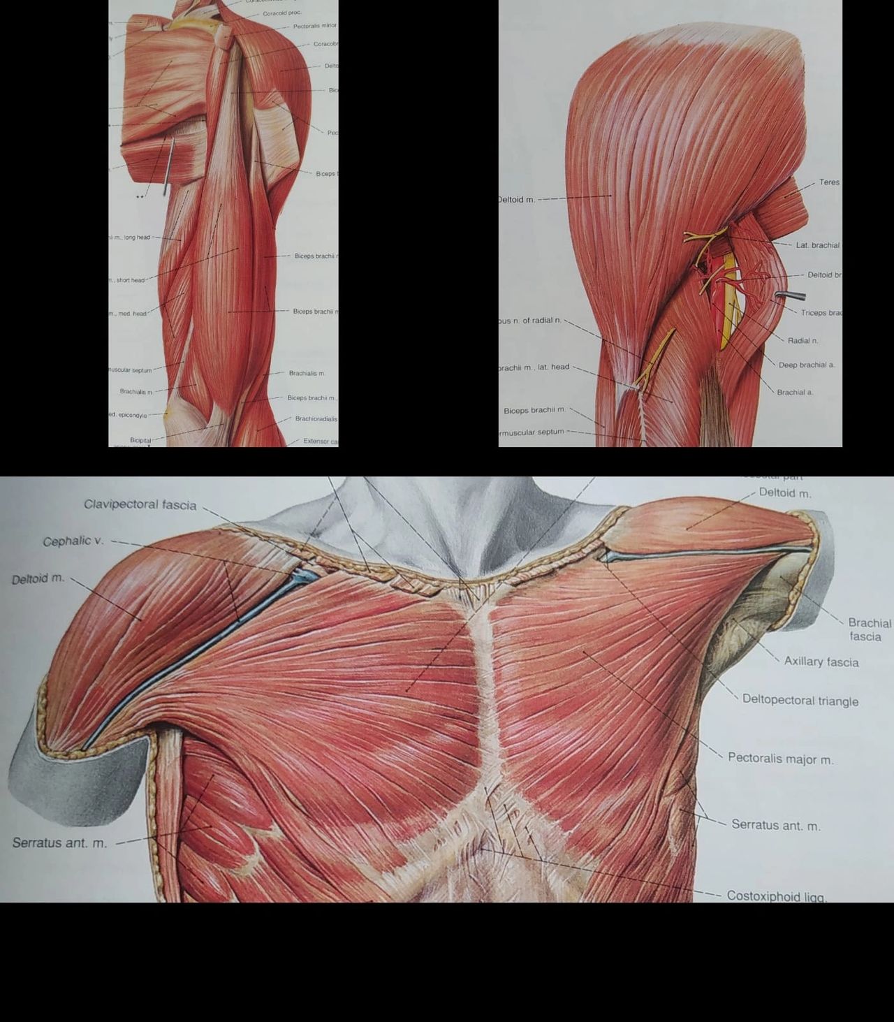
As mentioned above, every muscle can perform more than one movement, and every movement consists of more than one muscle. There are three main optional muscles that may interfere with flexion of the shoulder: Biceps Brachii, Pectoralis Major, and Anterior Deltoid.
While the Biceps Brachii is most commonly known for flexion of the elbow, it is important to note that both proximal insertions of the muscle are found on the Scapula. Due to their location, they are also taking part with the flexion of the shoulder.
Pectoralis Major, is also a big cause of discomfort. It’s a vast muscle that is involved in different movements in the shoulder. The Pectoralis Major originates from the Clavicle, Sternum and ribs, and attaches on the Humerus. It will pop with other movements too.
The last one of the three, is the Deltoid, specifically the anterior portion of it (hence the name Anterior Deltoid). The Deltoid divides into 3 main groups, of which we will discuss later on, and in accordance to the group’s location, so does the function they supply. The Anterior Deltoid is involved with shoulder flexion, and so is taken into consideration while checking the pain arising from the motion.
Shoulder Extension
Again, we will be looking at three main muscles: Long Head of Triceps, Latissimus Dorsi and Posterior Deltoid.
Unlike the Biceps Brachii, the Triceps consists of three heads, but only one of which crosses the shoulder and takes part of the movement. This head is called the Long Head of Triceps, and is the only part to take part of shoulder extension.
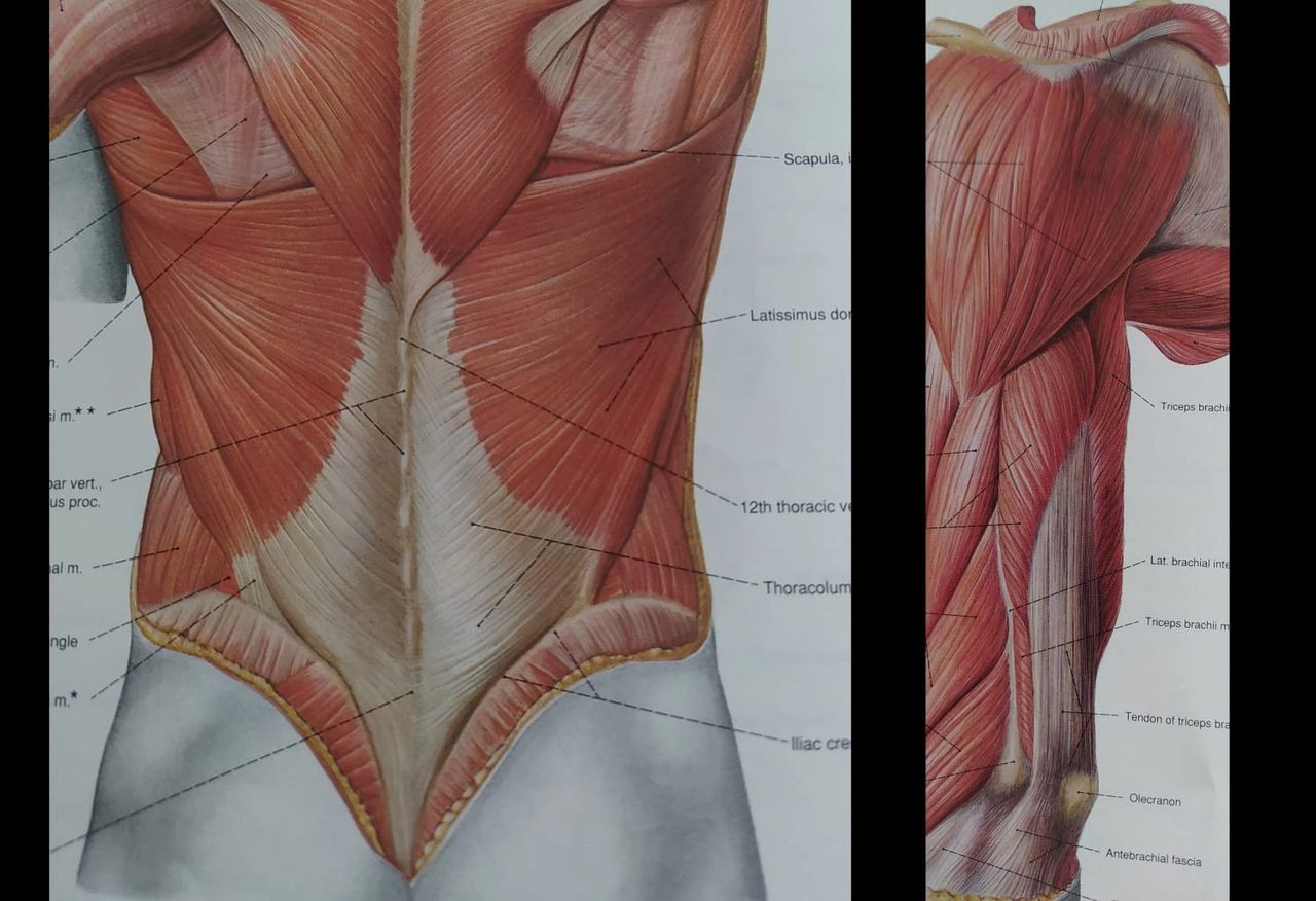
Next We’d have the Latissimus Dorsi. Often looked upon as the opposite muscle to the Pectoralis Major, the Latissimus Dorsi is vast, originating from the Thoracolumbar fascia and inserts into the Humerus.
As with flexion, the last of the three is the Deltoid, however, this time we are looking at the Posterior Deltoid.
Shoulder Abduction
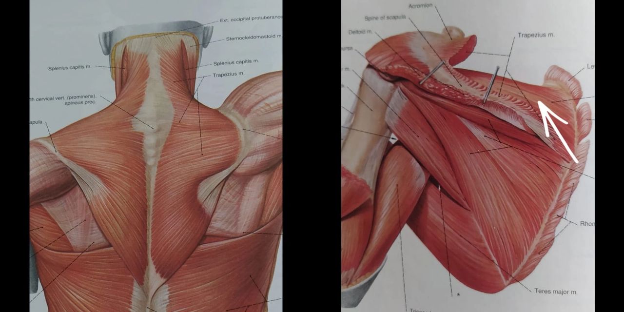
While it is a swift motion all the way through, it is divided into three muscles that are doing it: Supraspinatus for the first ~30 degrees, Middle Deltoid for 30 ~ 90, and Trapezius for the rest.
The first to start the motion is Supraspinatus, while it is part of the notorious Rotator Cuff muscles, it also plays an important role in abducting the shoulder, and bringing the Humerus into working range for the Deltoid to take from.
Once Deltoid takes over, it is the middle portion of it, and so the tests would be to the Middle Deltoid.
Once the Deltoid fully contract, the Trapezius will take over the rest of the motion, by rotating the Scapula upwards and bringing the whole arm above the head.
Shoulder Adduction
The major muscles in this case would be 2 muscles we have already examined: Pectoralis Major and Latissimus Dorsi.
Depending on the angle in which the shoulder is being adducted, one of them will be the prime mover. If the shoulder is being adducted to the front of the body, we will examine Pectoralis Major. However, if the shoulder is being adducted to the back the Latissimus Dorsi will be examined.
Shoulder Horizontal Abduction
Again, a muscle we have already looked at, for the horizontal abduction, the examination would focus on Posterior Deltoid. While there are other muscles that may be involved in the motion, they would require a more thorough examination of the movement itself.
Shoulder Horizontal Adduction
We have two prime muscles taking part of this movement, Anterior Deltoid and Pectoralis Major. This is where all the past motions we’ve discussed will come to play, as we will be able to differentiate which of them might be the cause of the pain.
Internal & External Shoulder Rotation
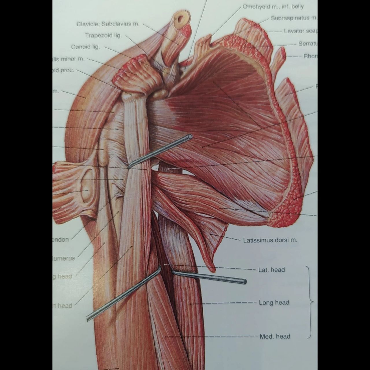
I’ve decided to combine those motions along as they are caused by the same group of muscles, the Rotator Cuff (Supraspinatus, Infraspinatus, Subscapularis and Teres Minor). External rotation is created by Supraspinatus, Infraspinatus and Teres Minor (refer to the image of Supraspinatus. Infraspinatus is right below it and Teres Minor pops below and a bit to the left). Which leaves Subscapularis to perform internal rotation.
While the above are the main muscles responsible for shoulder rotation, they are not the only ones.
nternal Rotation is also performed by Pectoralis Major, while the External Rotation also relies on Latissimus Dorsi.
Please Note that while pain can occur due to the performing muscle being tight, it is highly likely that the pain is caused from a resting muscle not allowing the motion to occur fluently, and every case should be examined as a stand alone case by a qualified practitioner.
The described above, are the main muscles that could be causing discomfort and pain. There could be more cases that would require further examination and tests. If you feel any issue with your shoulder, please go see a qualified therapist or your GP. For any questions you have, or if you wish to book an appointment with me, follow the links. Stay Safe!
Content Last Reviewed & Updated on September 2025 – All statistics, references, and therapy recommendations have been reviewed to ensure current best practices.
References
Images taken from Sobotta – Atlas of Human Anatomy, 11th English Edition, Vol. 1 & 2